Pulmonary Edema: Causes, Symptoms, and Preventive Measures
Imagine a world where understanding pulmonary edema isn't a daunting task? We're here to break down this complex issue, explain its causes, symptoms, diagnosis, and preventive measures in simple terms.

Are you living in fear of pulmonary edema? The constant worry about this dangerous condition can leave you feeling breathless. But imagine a world where understanding pulmonary edema isn't a daunting task. We're here to break down this complex issue, explain its causes, symptoms, diagnosis, and preventive measures in simple terms. With a little knowledge, you can tackle the fear of pulmonary edema head-on, promoting peace of mind and healthier outcomes. Remember - the first step to combating any health issue is understanding it.
What is Pulmonary Edema?
Pulmonary edema refers to the accumulation of excessive fluid within the lung's interstitial spaces or alveoli. This build-up hampers normal gas exchange between the air sacs (alveoli) and small blood vessels (capillaries) in the lungs. As a consequence, oxygen absorption becomes impaired while carbon dioxide elimination is compromised. Thus, pulmonary edema severely compromises respiratory function. The underlying pathophysiology involves an imbalance between fluid filtration into the lung tissues and subsequent removal by lymphatics. Fluid extravasation may occur due to increased hydrostatic pressure within pulmonary capillaries or increased permeability of the alveolar-capillary membrane caused by inflammation or injury. The excess fluid accumulates in interstitial spaces or fills up alveoli directly impairing their ability to effectively exchange gases.
FDA Disclaimer: The information contained in this document is for educational and informational purposes only, and is not intended as health or medical advice. Always consult a physician or other qualified health provider regarding any questions you may have about a medical condition or health objectives.

- 🌿 FIGHT WATER RETENTION - Bid farewell to bloating and puffiness with the assistance of these water pills! This powerful diuretic is suitable for both men and women, effectively eliminating excess fluids from your system.
- 🌿 GENTLE & FAST RELIEF - Restore your water balance in a holistic manner. THese water weight pills stimulate increased urination, providing relief from swelling caused by edema in your feet, ankles, and legs.
- Affiliate Disclosure: If a product is purchased through one of our affiliate links, we may receive a commission at no extra cost to you. This helps to support our blog 😄
Importance of Understanding Pulmonary Edema
Comprehending pulmonary edema is crucial for several reasons. Firstly, it can arise as a result of numerous medical conditions such as heart failure, myocardial infarction (heart attack), acute respiratory distress syndrome (ARDS), high altitude exposure, drug toxicity, or even severe infections like pneumonia. Understanding these underlying causes assists clinicians in identifying potential risk factors amongst patients presenting with symptoms that align with pulmonary edema.
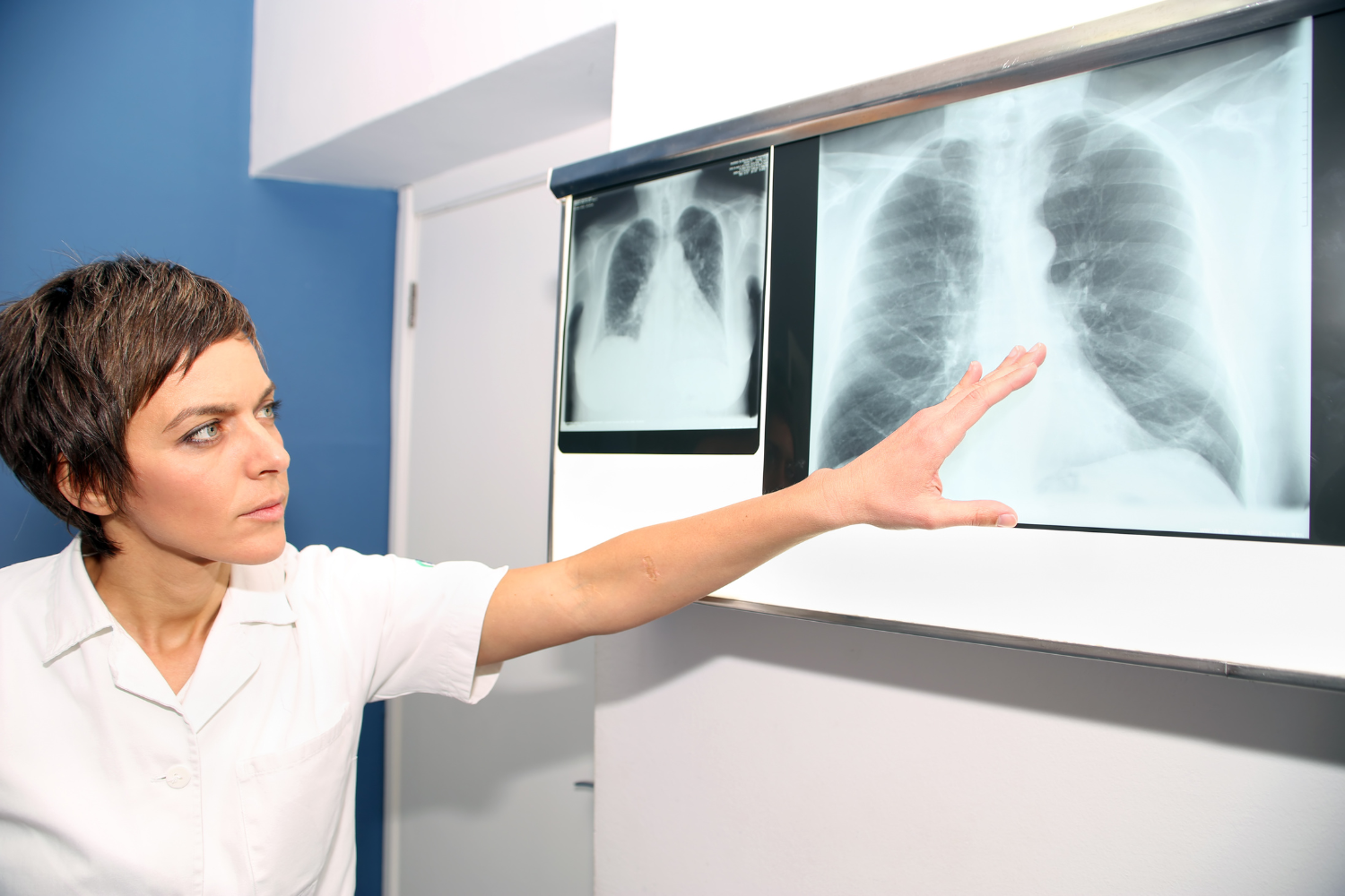
Secondly, recognizing signs and symptoms of pulmonary edema is imperative for timely diagnosis and appropriate management. Symptoms can range from mild dyspnea (shortness of breath) to severe respiratory distress, coughing up frothy pink-tinged sputum, fatigue, weakness, and even chest pain or discomfort. By being familiar with these clinical manifestations, individuals can seek medical attention promptly, thereby increasing their chances of receiving timely treatment. Understanding the diagnostic approach for pulmonary edema aids in accurate assessment and differentiation from other conditions with similar presentations. Diagnostic modalities such as physical examination findings (e.g., increased respiratory rate and effort, lung auscultation revealing crackles or wheezing), imaging studies like chest X-rays (showing bilateral infiltrates and cardiomegaly), echocardiography (assessing cardiac function), as well as laboratory tests including complete blood count, electrolyte panel, and B-type natriuretic peptide levels are crucial for confirming the diagnosis.

What Causes Pulmonary Edema?
Causes and Risk Factors
- Acute Respiratory Distress Syndrome (ARDS): This involves severe inflammation and damage to the lung tissue. It increases the permeability of the alveolar-capillary membrane, causing fluid to leak into the air sacs—leading to pulmonary edema.
- Exposure to High Altitudes: Rapid exposure to decreased oxygen levels at higher altitudes may trigger High Altitude Pulmonary Edema (HAPE). This condition results from the constriction of blood vessels in the lungs, followed by leakage of fluid into lung tissues.
- Age: Advanced age is associated with decreased cardiovascular function and increased susceptibility to heart failure or other cardiac conditions that can lead to pulmonary edema.
- Smoking History: Smoking damages both lung tissue and blood vessels over time, making smokers more susceptible to the condition.
- Hypertension (High Blood Pressure): High blood pressure puts strain on blood vessels throughout the body, including those within the lungs. This can increase hydrostatic pressure within the pulmonary capillaries and contribute to pulmonary edema.
- Pre-existing Heart or Kidney Disorders: Individuals with pre-existing heart or kidney disorders are at higher risk due to the underlying pathophysiological processes that affect fluid balance within the body.
What is Flash Pulmonary Edema?
For those interested, flash pulmonary edema is a rapid-onset condition typically caused by acute left ventricular failure. This sudden failure can result from several underlying conditions, including:
- Acute Myocardial Infarction: An abrupt blockage of one or more of the coronary arteries can cause a heart attack, subsequently leading to flash pulmonary edema.
- Rapid-Onset Hypertension: Sudden, severe high blood pressure can strain the heart and lead to left ventricular failure, triggering flash pulmonary edema.
- Mitral Valve Dysfunction: Issues with the mitral valve, such as mitral valve prolapse or stenosis, can impede the flow of blood from the left atrium to the left ventricle, potentially causing flash pulmonary edema.
- Renal Artery Stenosis: This condition, characterized by narrowing of the arteries that carry blood to the kidneys, can lead to refractory hypertension and subsequently flash pulmonary edema.
This is not an exhaustive list, and other medical conditions can also result in flash pulmonary edema. Always consult a healthcare professional for a comprehensive understanding of your risk factors.
Pathophysiology of Pulmonary Edema
The pathophysiological mechanisms underlying pulmonary edema involve a disruption in the delicate balance between hydrostatic pressure, permeability of the alveolar-capillary membrane, and lymphatic drainage. Normally, hydrostatic pressure within the pulmonary capillaries favorably promotes fluid movement outwards into surrounding tissues. However, in pulmonary edema, there is an excessive increase in hydrostatic pressure. This can be caused by cardiac issues such as heart failure or myocardial infarction, which impair the heart's ability to efficiently pump blood forward. The increased pressure within these vessels forces fluid out and into the lung tissues. In addition to elevated hydrostatic pressure, changes in the permeability of the alveolar-capillary membrane contribute significantly to pulmonary edema. Under normal circumstances, this membrane acts as a barrier preventing fluid from leaking into the air sacs. However, when inflammation or damage occurs due to conditions like ARDS or infection, it disrupts this barrier and increases its permeability. This allows more fluid to pass through and accumulate within lung tissues.
Note: We are an affiliate of products we feel are helpful to our audience. By no means is this medical advice and please enlighten yourself with knowledge and check out the products we link to. Always consult a healthcare professional with questions and curiosity on the subjects we cover.
Furthermore, impaired lymphatic drainage exacerbates fluid accumulation in cases of pulmonary edema. The lymphatic system plays a crucial role in maintaining fluid balance throughout the body by collecting excess interstitial fluid and returning it to circulation. In instances where lymphatic vessels are compromised or overwhelmed by excessive fluid volume (as seen in conditions like heart failure), proper drainage may be hindered. Consequently, interstitial fluids build up within lung tissues leading to further development of pulmonary edema. Understanding these intricate pathophysiological mechanisms associated with increased hydrostatic pressure, altered membrane permeability, and impaired lymphatic drainage is vital for comprehending the development and progression of pulmonary edema. By addressing these underlying factors, healthcare professionals can effectively tailor treatment strategies to manage this potentially life-threatening condition.
Clinical Presentation and Symptoms
Acute vs Chronic Pulmonary Edema
When it comes to pulmonary edema, it is crucial to distinguish between acute and chronic presentations as they have distinct clinical characteristics and implications for management. Acute pulmonary edema typically occurs suddenly and is often associated with a severe underlying condition such as myocardial infarction or heart failure exacerbation. On the other hand, chronic pulmonary edema develops gradually due to long-standing heart or lung diseases, such as congestive heart failure or chronic obstructive pulmonary disease (COPD). Recognizing the time frame of symptom onset is vital in determining appropriate treatment strategies.
Common Symptoms
Pulmonary edema manifests through a range of distressing symptoms that can significantly impact an individual's quality of life. The most common symptom experienced by patients with pulmonary edema is dyspnea, which refers to difficulty breathing or shortness of breath. This sensation may worsen when lying flat (orthopnea) or during physical exertion (paroxysmal nocturnal dyspnea). Additionally, coughing with frothy sputum is frequently observed in individuals with pulmonary edema. The sputum may appear pinkish due to the presence of blood, indicating further respiratory compromise.
Dyspnea (Shortness of Breath)
Dyspnea is the hallmark symptom of pulmonary edema and results from impaired oxygen exchange within the lungs. Patients often describe a sensation of suffocation or air hunger that leads to significant discomfort and anxiety. Dyspnea may be accompanied by rapid shallow breathing (tachypnea), as the body attempts to compensate for inadequate oxygenation. It is important to note that dyspnea severity can vary depending on the extent of fluid accumulation in the lungs and underlying causative factors.
Coughing with Frothy Sputum
Coughing with frothy sputum is another characteristic symptom of pulmonary edema. As fluid accumulates in the lungs, it mixes with air and leads to the production of foamy or pinkish sputum. This frothy appearance reflects increased capillary permeability and leakage of fluid into the alveoli. The cough may become persistent and interfere with daily activities, causing further distress for individuals suffering from pulmonary edema.
Fatigue and Weakness
Fatigue and weakness are frequently reported symptoms in patients with pulmonary edema. The compromised oxygen exchange resulting from fluid buildup in the lungs can lead to reduced oxygen delivery to various organs, including skeletal muscles. This inadequate oxygen supply can cause muscle fatigue and weakness as these tissues struggle to perform their normal functions. Furthermore, systemic effects related to heart failure or other underlying conditions may contribute to overall fatigue experienced by individuals affected by pulmonary edema.
Chest Pain or Discomfort
Chest pain or discomfort is another symptom that can be associated with pulmonary edema, particularly when caused by cardiac etiologies such as myocardial infarction or unstable angina. While chest pain may indicate other conditions, its presence alongside respiratory distress should raise suspicion for concurrent pulmonary involvement. This pain is typically described as a pressure-like sensation in the chest that may radiate to other areas such as the neck, jaw, back, or arms. Recognizing the clinical presentation and symptoms of pulmonary edema is essential for early detection and appropriate management. Acute versus chronic delineation aids in understanding disease progression while guiding treatment decisions accordingly. Common symptoms like dyspnea (shortness of breath), coughing with frothy sputum, fatigue and weakness, along with chest pain or discomfort serve as vital indicators warranting prompt medical attention to mitigate potential complications associated with this serious condition.
Diagnosis and Evaluation
Physical examination findings
During the evaluation of a patient suspected of having pulmonary edema, the physical examination plays a vital role in confirming the diagnosis. Several findings can provide valuable insights into the presence and severity of pulmonary edema. One common physical finding is an increased respiratory rate and effort. Patients may exhibit rapid, shallow breathing due to an inability to adequately oxygenate their blood. Additionally, they may demonstrate signs of respiratory distress, such as retractions or flaring of nostrils. Another noteworthy finding is the presence of crackles or wheezing on lung auscultation. Crackles are discontinuous, high-pitched sounds that occur during inspiration and result from the sudden opening of closed airways due to fluid accumulation in alveoli. In contrast, wheezing is a continuous musical sound occurring during expiration, suggesting airway constriction secondary to pulmonary edema. Moreover, peripheral edema can often be observed during physical examination as a consequence of fluid retention seen commonly in patients with heart failure-induced pulmonary edema. The swelling usually affects dependent areas such as lower extremities but can also extend to other parts like sacrum or hands.
Imaging studies
Imaging studies are crucial for confirming the diagnosis and assessing the extent of pulmonary edema. Chest X-ray is one commonly employed modality that provides valuable information about lung parenchymal changes associated with this condition. Bilateral infiltrates are typically seen on chest X-ray due to fluid accumulation within alveoli, giving rise to a hazy appearance throughout both lungs. Furthermore, cardiomegaly may be evident on chest X-ray as an enlarged cardiac silhouette resulting from underlying heart failure contributing to pulmonary congestion leading to edema formation. In addition to chest X-rays, echocardiography serves as another important imaging tool in evaluating patients with suspected pulmonary edema. Echocardiography allows for the assessment of cardiac anatomy and function, helping to identify any structural abnormalities or impaired ventricular contractility that may be contributing to the development of pulmonary edema.
My Recommendation to Gain a Further Understanding On Pulmonary Edema
As a writer and blogger, I find it important to share resources I think are worth the read. The intricacies of disease and the diverse range of underlying causes highlight the importance of thorough knowledge in navigating and managing this health challenge. Increased awareness and comprehension of pulmonary edema can empower individuals to consciously avoid illness and pursue proactive healthcare measures. To delve deeper into this subject, consider reading Noninvasive Ventilation in Medicine: Clinical and Pulmonary Critical Care. This comprehensive resource offers a wealth of information that can be instrumental in understanding pulmonary edema and its implications. This copy also has some clinical trial information for those are interested in that. I've also attached a picture below for more interest:
Noninvasive Ventilation in Sleep Medicine and Pulmonary Critical Care
1st Edition, 2020
Affiliate Disclosure: This document contains affiliate links to products. We may receive a commission for purchases made through these links, at no extra cost to you. It helps us to make more great content for our audience and support the newsletter.
Key Takeaways on Pulmonary Edema
- Understand the Basics: Pulmonary edema is a condition in which fluid accumulates in the lungs, making breathing difficult and potentially leading to respiratory failure.
- Know the Causes: Various medical conditions can trigger pulmonary edema, including heart conditions like mitral valve stenosis or systemic diseases like renal artery stenosis.
- Be Aware of the Risks: Not everyone exposed to the causes of pulmonary edema will develop the condition, but certain factors increase risk, such as pre-existing cardiovascular disease or kidney issues.
- Recognize the Symptoms: Shortness of breath, especially when lying down; feeling of "drowning" or suffocating; wheezing or gasping for breath; anxiety or restlessness; and coughing up frothy, pink-tinged mucus are common symptoms.
- Seek Immediate Medical Attention: If you or someone else experiences symptoms of pulmonary edema, seek emergency medical care. It's a serious condition that can rapidly become life-threatening.
- Embrace Prevention: Lifestyle changes and medication can help prevent pulmonary edema, especially if you have a condition that puts you at higher risk.
- Explore Further: Books such as "Noninvasive Ventilation in Medicine: Clinical and Pulmonary Critical Care" offer comprehensive information on this complex disease.
FDA Disclaimer: The information contained in this document is for educational and informational purposes only, and is not intended as health or medical advice. Always consult a physician or other qualified health provider regarding any questions you may have about a medical condition or health objectives.